Edinburgh Imaging

Diffusion MR & tractography DTI

Diffusion MRI (dMRI) measures the mobility of water molecules in the brain.
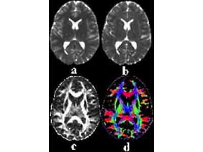
Diffusion tensor MR image.
Standard axial diffusion tensor MR (DT-MR) parametric maps obtained from a healthy volunteer. (a) T2W image of brain (grey) & cerebrospinal fluid (white). (b) Magnitude of water molecule diffusion (mean diffusivity (MD)). (c) Scalar measure of diffusion anisotropy (FA), i.e. the deviation from pure isotropic diffusion of water mobility in vivo. This diffusion anisotropy arises due to the presence of axonal membranes & myelin which causes water molecules to diffuse preferentially along the long axes of ordered axonal fibres rather than across them; these DT parameters are therefore thought to provide useful biomarkers of WM fibre tract integrity, with low values of (grey) & high values of (white) FA indicating intact healthy coherent axons. (d) Simple colour composite map of the principal direction of water mobility (red = left / right; green = anterior / posterior; blue = superior / inferior).

 Water molecules tend to diffuse along, rather than across, brain white matter fibres.

Consequently, it is possible to map the structure & integrity of major water matter tracts in 3 dimensions, using Diffusion Tensor Imaging (DTI Tractography).

Mapping the structure of white matter tracts, effectively the brain's wiring, in vivo, allows us to understand how different brain regions are connected & how diseases such as stroke, intracranial tumours and schizophrenia affect white matter & cause neurological problems.

The video clip is of a three dimensional rotating colour coded map illustrating the white matter tracts produced from DT-MRI data acquired at the Edinburgh Imaging Facility WGH


Contact

If you wish further information on any of the above activities, then please contact Dr Mark Bastin.